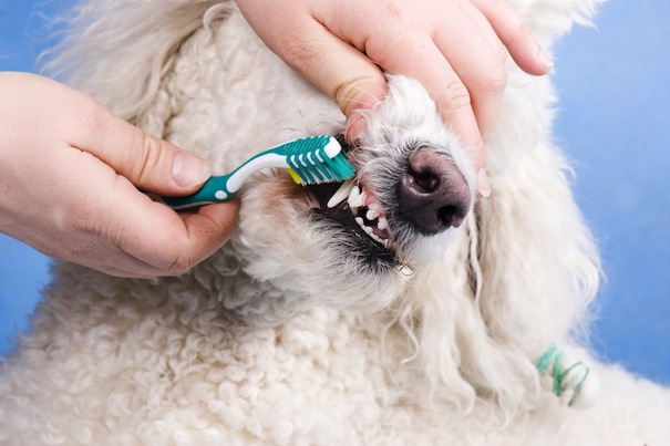
Much like an iceberg, the visible area of our pet’s teeth (or crown) is only the tip! At least 50% of the pet’s tooth is hidden below the gum line.
Dental disease usually begins with a build-up on the teeth which hardens to form plaque. This plaque then leads to inflamed gums – called gingivitis – which progresses to erosion of the gums and pocketing, where plaque migrates below the gum line, causing advanced dental issues such as tooth loss, painful abscesses and general infection. These can lead to a decline in your pet’s health and wellbeing. In severe cases, these harmful oral bacteria can spread throughout the body and cause issues with the animal’s heart.
A staggering 80% of pets will suffer from some form of dental disease before the age of five. Early indicators are bad breath, trouble chewing biscuits or treats, visible tartar and sore or inflamed gums.
We can address the visible problems by keeping on top of tooth brushing and providing dental chews. However, seeing below the gum line to visualise the damage caused by advancing plaque and bacteria requires more specialist equipment.
Dental x-ray has been commonplace in human dentistry for many years; however, this is still an emerging technology in veterinary practice. This allows us to visualise below the gum line to see the 50% of the tooth which is normally hidden to us.
Dental x-ray generators allow us to pinpoint diseased teeth and select the most time-appropriate treatment to save pets from unnecessary time under anaesthetic. We can then see which teeth require extraction and which have already started the resorption process and are being absorbed by the body. Dental x-rays can be performed very quickly as part of a routine dental procedure carried out under anaesthetic – ask your veterinary practice if they are trained to offer this service, as our clinicians are, and make sure you stay on top of your pet’s dental health.
Jo Wright DipAVN RVN
Severn Edge Vets


